The structure of mouse RIPK1 RHIM-containing domain as a homo-amyloid and in RIPK1/RIPK3 complex
Solid-state NMR reveals the amyloid architecture of mouse RIPK1 and structural changes in RIPK1/RIPK3 fibrils.
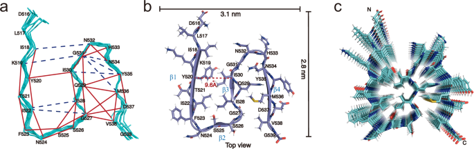
Fig. 5 · Liu et al., Nature Communications (2024) · CC BY-NC-ND 4.0
Research summary
Using solid-state NMR, this study resolves an N-shaped, four-strand amyloid fold in the mouse RIPK1 RHIM-containing region. Comparisons with RIPK3 and mixed RIPK1/RIPK3 fibrils reveal differences in structure and rigidity, providing insight into RHIM-mediated interactions in necroptosis.
Citation
Liu, J., Wu, X.-l., Zhang, J., Li, B., Wang, H.-y., Wang, J. & Lu, J.-x. (2024). The structure of mouse RIPK1 RHIM-containing domain as a homo-amyloid and in RIPK1/RIPK3 complex. Nature Communications, 15, 6975. https://doi.org/10.1038/s41467-024-51303-y
Figure credit
Figure 5 is reproduced without modification from Liu et al., Nature Communications (2024). © The Author(s) 2024. Licensed under Creative Commons Attribution–NonCommercial–NoDerivatives 4.0 International.